Neuerungen in der Nomenklatur der Pilze
Ätiologie, Pathogenese und Prävalenz
Klinische Symptomatik
Die Dermatophytose ist eine kutane Infektion, die durch mehrere keratophile Pilzarten (Dermatophyten) verursacht wird. Sie stellt eine ernstzunehmende sowie häufig auftretende kontagiöse Hautkrankheit bei Hunden und Katzen dar. Die Bedeutung dieser Krankheit für Tierbesitzer basiert auf ihrem zoonotischen Potenzial. Ihre Prävalenz variiert mit dem Klima und der lokalen Dermatophytenprävalenz. Die häufigsten Infektionen bei Hund und Katze erfolgen durch die Genera Microsporum (M.), Nannizzia (N.) oder Trichophyton (T.). Ziel dieses Artikels ist eine Zusammenfassung der Neuerungen hinsichtlich Taxonomie, Diagnose und Therapieempfehlungen sowie der aktuell überarbeiteten Empfehlungen der World Association of Veterinary Dermatology.
Neuerungen in der Nomenklatur der Pilze
Die Nomenklatur der Pilze entwickelt sich stetig weiter [101]. Mykologen sind darum bemüht, die Zugehörigkeit der Pilze phylogenetisch korrekt zu klassifizieren [101] und eine einheitliche Nomenklatur für Pilze zu schaffen [16][43][44][101]. Grundsätzlich wird nach molekularen Analysen eine Zusammenführung der beiden Klassifizierungssysteme, der sexuellen Eumycota und der asexuellen Deuteromycota, angestrebt [32]. Bislang wurden häufig sexuelle und asexuelle Stadien ein- und desselben Pilzes als unterschiedliche Pilze identifiziert. Eine neue phylogenetische Studie hat die Taxonomie der Dermatophyten hinsichtlich ihres Hintergrunds überprüft und Umgruppierungen vorgenommen [28]. Arthroderma enthält jetzt 21 Arten [28], während sich die Gattung Microsporum auf 3 Arten beschränkt: M. audouinii, M. canis und M. ferrugineum. Die verbleibenden geophilen und zoophilen Arten, die zuvor als Microsporum -Subspezies galten, wurden auf die Gattungen Lophophyton (L.), Nannizzia (N.) und Paraphyton (P.) übertragen [28]. Epidermophyton enthält nun 1 Art, Ctenomyces 1 Art und die Gattung Lophophyton wurde ebenfalls auf eine einzige Art reduziert: Lophophyton gallinae. Nannizzia umfasst nun 9 Arten. N. gypsea, N. persicolor, N. fulva sowie N. nana gehörten zuvor zur Gattung Microsporum. Trichophyton beeinhaltet 16 Arten. T. ajelloi und T. terrestre wurden nun der Gattung Arthroderma zugeordnet [28]. Der T. mentagrophytes -Komplex besteht aktuell aus anthropophilen und zoophilen Spezies (siehe Umgruppierung Tab. 2) [32][76][100]. In diesem Artikel wird folglich M. gypseum als N. gypsea bezeichnet (Tab. 1).
Ätiologie, Pathogenese und Prävalenz
Dermatophyten sind keratinophile Pilze, die Haare, Nägel oder Krallen infizieren [6] und oberflächliche Mykosen verursachen können [5] (Abb. 1). Per definitionem wird die Dermatophytose bei Hunden und Katzen durch eine oberflächliche Infektion der keratinisierten Hautstrukturen durch geophile, zoophile oder anthropophile Pilze ausgelöst [66]. Die Übertragung erfolgt durch direkten Kontakt mit infektiösen, fragmentierten Pilzhyphen, sog. Arthrosporen [5][6], die anschließend dem Stratum corneum anhaften [6]. Durch Virulenzfaktoren wie Keratinasen und Proteasen bahnt sich der Pilz seinen Weg in die Epidermis [5][6], begünstigt durch eventuelle Mikrotraumen der Epidermis. Ansteckungsmöglichkeiten sind sowohl Artgenossen als auch Haustierzubehör oder Ektoparasiten [76][85].

Abb. 1: Makrokonidien von Microsporum canis , H & E-Färbung, Vergrößerung 1000x.
Aufgrund der vielseitigen klinischen Symptomatik ist die Dermatophytose eine häufige Differenzialdiagnose in der dermatologischen Sprechstunde. Das zoonotische Potenzial [84] erhöht noch die Bedeutung der Krankheit. Die anerkannte Zoonose verursacht bei Menschen Hautläsionen, die bei immunkompetenten Individuen in der Regel gut abheilen [76], während sich immunsupprimierte Menschen oft einer länger dauernden Behandlung unterziehen müssen [9]. Bei Mensch und Tier sind mehr als 30 verschiedene Hautpilzspezies beschrieben [106].
Als häufigste Erreger treten bei Haustieren M. canis, N. gypsea sowie T. mentagrophytes auf [76]. Für die Nomenklatur wird, basierend auf ihrem natürlichen Lebensraum, eine Unterteilung in zoophile, anthropophile und geophile Spezies vorgenommen [66]. Zu den zoophilen Genera, die auf tierischen Wirten leben und sich vornehmlich vom Keratin der Haut ernähren, gehören M. canis (betroffen sind am häufigsten Katzen und Hunde), mehrere Spezies des T. mentagrophytes -Komplexes (Nager, Hasenartige, Igel), T. verrucosum (Rinder), N. persicolor (Vorkommen überwiegend auf Wühlmäusen), N. nana (Schwein), T. equinum (Pferd) sowie M. equinum (vornehmlich bei Pferden zu isolieren) [66]. N. gypsea ist der am häufigsten bei Hunden und Katzen isolierte geophile Pilz [76]. Vertreter der geophilen Gattungen ernähren sich vorwiegend von Keratin aus Horn, Haar und Federn, die im oder auf dem Erdreich verwesen, anstatt wie zoophile und anthropophile Arten diese Nährstoffquellen auf dem Wirt zu verstoffwechseln [66]. Die meisten dieser Pilze sind nicht pathogen für Mensch und Tier, doch kann es zu sporadischen Infektionen mit geophilen Pilzen durch kontaminierte Erde kommen [76].
Studien zur physiologischen Pilzflora von gesunden Katzen und Hunden haben gezeigt, dass M. canis nicht zum physiologischen Hautmikrobiom von Hunden oder Katzen gehört [48][62][63][67][68] . Bei Hunden wurden in mehreren Studien Alternaria und Cladosporium als häufigste physiologische Pilzarten der Hautflora isoliert. Die physiologische Pilzflora von Hauskatzen ist hingegen sehr vielfältig [68]. In Bezug auf Pilze entspricht das kutane Mikrobiom bei Hund und Katze am ehesten der Umgebungspilzflora. Moriello et al. [68] konnten bei der Untersuchung von Katzenhaaren bis zu 13 Saprophyten und 2 Dermatophyten isolieren, wobei Penicillium, Aspergillus, Cladosporium spp. und Alternaria die am häufigsten isolierten Arten waren.
Die Prävalenz von Dermatophytosen ermittelte eine kanadische Studie [95] mit 0,71 % beim Hund und 3,6 % bei der Katze. In einer Studie aus Großbritannien [46] betrug sie 0,53 % beim Hund und 1,3 % bei der Katze. In Europa ist M. canis ist für 50–90 % der kaninen und felinen Dermatophytosen verantwortlich [13][14][36][99]. M. canis wird hauptsächlich durch Katzen übertragen [59], N. gypsea durch kontaminierte Böden und Trichophyton sp. durch Nagetiere als Vektoren [76][85]. Die Katze ist grundsätzlich das wichtigste Reservoir für M. canis [59]. Andere Dermatophyten haben bei unseren Haustieren eine weitaus geringere Prävalenz [59].
Juvenile, in dicht besiedelten Gebieten lebende Wildtiere sowie Tiere an warmen Standorten haben ein höheres Infektionsrisiko [76][91]. Durch den Kontakt mit kontaminiertem Boden scheinen Jagd- und Arbeitshunde (z. B. wie Groenendael, Foxterrier, Labrador Retriever, Deutsch Kurzhaar, Belgier, Deutscher Schäferhund, Jagdterrier, Beagle, Jack Russell Terrier und Pointer) vor allem für N. gypsea und N. persicolor prädisponiert zu sein [11][19][80]. Yorkshire Terrier zeigen sich bei M. canis -Infektionen deutlich überrepräsentiert [1][3][10][15][21][76][112]. Katzen langhaariger Rassen, insbesondere Perserkatzen, werden als prädisponiert angesehen [55][95], allerdings sind Perserkatzen in klinischen Studien ohnehin überrepräsentiert [95]. Mehrere Studien beschreiben einen Zusammenhang zwischen Dermatophytose und Hyperadrenokortizismus bei Hunden [22][42][57][114]. Ferner existiert ein Artikel über Dermatophytose bei 6 Yorkshire Terriern mit unterschiedlichen Begleiterkrankungen (Leishmaniose: n = 4, Ehrlichiose: n = 1, Diabetes mellitus: n = 1) [21]. Katzen mit einer FIV- oder FeLV-Infektion weisen zwar eine größere Vielfalt an saprophytären Pilzorganismen auf, sind aber nicht vermehrt Träger von Dermatophyten [58][65][97].
Klinische Symptomatik
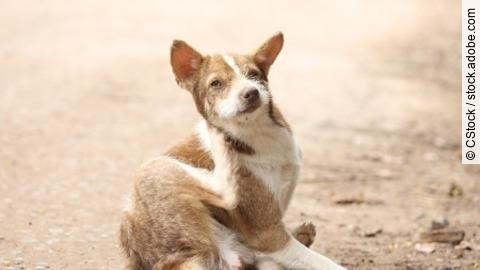
Beim Hund äußert sich die Erkrankung vor allem durch alopezische Läsionen, Schuppen, Krusten, Erytheme, follikuläre Papeln, Follikulitis und Hyperpigmentierung [66]. In der Regel besteht kein bis mäßiger Juckreiz [53]. Grundsätzlich kommt bei nicht juckenden hypotrichoten Läsionen eine Dermatophytose differenzialdiagnostisch infrage [32]. Dermatophytenläsionen liegen klassischerweise asymmetrisch vor [76]. Selbst zugefügte Läsionen können sekundär bakteriell infiziert sein [53]. Läsionen im Gesichts- und Nasenbereich finden sich vor allem bei Arbeitshunden [53]. Eine Beteiligung des Krallenbetts ist selten [53] und in Einzelfällen kommt es auch zu Veränderungen des Krallenwachstums (Onychogryphose) [66]. Des Weiteren kann beim Hund eine selten beschriebene pustulöse Dermatophytose auftreten, die dem Pemphigus foliaceus ähnlich erscheint [90].
Katzen erkranken hauptsächlich durch M. canis [53][72] und der Pilz verursacht meist nur relativ milde Läsionen mit geringen Entzündungsreaktionen [53] (Abb. 4). Vorwiegend treten auch bei dieser Spezies Haarausfall, Krusten, Papeln, Schuppen, Erythem sowie Hyperpigmentierung auf und der Juckreiz stellt ein variables Symptom dar [66]. Die Läsionen zeigen sich am häufigsten im Gesichtsbereich, an den Ohren und der Mundpartie [31][72]. Im Verlauf der Erkrankung kann die Dermatophytose beim Putzakt auf Pfoten und andere Körperregionen übertragen werden [31][72]. Differenzialdiagnostisch kommen vor allem bakterielle Infektionen, Autoimmunerkrankungen wie Alopezia areata und Ektoparasitosen, wie z. B. die Demodikose, infrage [21][53][91].

Abb. 4: Microsporum canis -Hautläsionen bei Katzen am Vorderbein (a) und Kopf (b). Auffällig sind die kreisrunden haarlosen Hautareale.
Bei Hunden und Katzen können selten Pseudomyzetome und Kerione auftreten [25][37]. Beim Kerion besteht eine hochgradige Entzündungsreaktion mit Furunkulose, die zu einer typischen erhabenen, alopezischen Schwellung von klassischerweise bis zu 5 cm im Durchmesser führt. Bei den Erregern handelt es am häufigsten um N. gypsea , T. mentagrophytes und M. canis . Das Zoonoserisiko wird geringer eingestuft als bei herkömmlichen Dermatophyteninfektionen. Eine Spontanremission ist zu erwarten, doch kann eine sekundäre bakterielle tiefe Pyodermie das klinische Bild beeinflussen. Eine Dermatophytenkultur hat nicht immer Aussagekraft, da die betroffenen Haarschäfte entweder fehlen oder in der Dermis rupturiert sind, wo sie wie Fremdkörper eine Entzündung fördern. In chronischen Fällen kommen differenzialdiagnostisch Neoplasien, atypische Mykobakterieninfektionen oder Pseudomyzetome infrage. Histologisch finden sich knotige Pyogranulome oder seltener Granulome in der Dermis, oft auch mit einer Beteiligung von Eosinophilen, in deren Zentren Reste von Haarschäften oder Sporen und Hyphen enthalten sein können. Allerdings lässt sich der Dermatophyt in stark entzündetem Gewebe manchmal nicht nachweisen [38].
Bei dermatophytischen Pseudomyzetomen handelt es sich um erythematöse, oft alopezische, erhabene, ulzerative und exsudative oder fistelnde Umfangsvermehrungen [25][37], die durch Dermatophyten wie beispielsweise T. mentagrophytes ausgelöst werden [38]. Histologisch zeigen sich Granulome mit Pilzaggregaten, die von Antigen-Antikörper-Komplexen umgeben (Splendore-Hoeppli-Reaktion) und mit Riesenzellen, Neutrophilen und Makrophagen infiltriert sind. Pseudomyzetome werden vor allem bei Perserkatzen, Yorkshire Terriern und Manchester Terriern beobachtet. Wahrscheinlich entstehen sie durch eine traumatische Implantation der Pilzorganismen in die Dermis. Die Gewebeknoten erreichen einen Durchmesser von 5–20 mm [38]. Sie können ulzerieren und griesförmiges bis seropurulentes Material freisetzen, das sich für die Dermatophytenkultur oder PCR nutzen lässt. Da die PCR auch tote Pilzsporen nachweist, ist zur Verifizierung eine Kultur oder histopathologische Untersuchung geeignet. Differenzialdiagnostisch kommen systemische Mykosen, Kryptokokkose, Sporotrichose, bakterielle Hautinfektionen und bakterielle Pseudomyzetome (verursacht durch Staphylococcus spp., Pseudomonas spp., Proteus spp. oder Streptococcus spp.) sowie Neoplasien infrage [38].
Der Originalartikel ist erschienen in:
1 Abramo F, Vercelli A, Mancianti F. Two cases of dermatophytic pseudomycetoma in the dog: an immunohistochemical study. Vet Dermatol 2001; 12: 203-207
2 Achten G. The use of detergents for direct mycologic examination. J Invest Dermatol 1956; 26: 389-397
3 Angarano DW, Scott DW. Use of ketoconazole in treatment of dermatophytosis in a dog. J Am Vet Med Assoc 1987; 190: 1433-1434
4 Asawanonda P, Taylor CR. Wood’s light in dermatology. Int J Dermatol 1999; 38: 801-807
5 Baldo A, Tabart J, Vermout S. et al. Secreted subtilisins of Microsporum canis are involved in adherence of arthroconidia to feline corneocytes. J Med Microbiol 2008; 57: 1152-1156
6 Baldo A, Monod M, Mathy A. et al. Mechanisms of skin adherence and invasion by dermatophytes. Mycoses 2012; 55: 218-223
7 Bayerl V. Prüfung der keimreduzierenden Wirkung eines Dampfreinigungsgerätes anhand mikrobiologischer Untersuchungen [Dissertation]. München: Ludwig-Maximilians-Universität; 2016
8 Bechter R, Schmid BP. Teratogenicity in vitro – a comparative study of four antimycotic drugs using the whole-embryo culture system. Toxicol In Vitro 1987; 1: 11-15
9 Berg JC, Hamacher KL, Roberts GD. Pseudomycetoma caused by Microsporum canis in an immunosuppressed patient: a case report and review of the literature. J Cutan Pathol 2007; 34: 431-434
10 Bergman RL, Medleau L, Hnilica K. et al. Dermatophyte granulomas caused by Trichophyton mentagrophytes in a dog. Vet Dermatol 2002; 13: 51-54
11 Bond R, Middleton DJ, Scarff DH. et al. Chronic dermatophytosis due to Microsporum persicolor infection in three dogs. J Small Anim Pract 1992; 33: 571-576
12 Bossche HV, Koymans L, Moereels H. P450 inhibitors of use in medical treatment: focus on mechanisms of action. Pharmacol Ther 1995; 67: 79-100
13 Breuer-Strosberg R. Reported frequency of dermatophytes in cats and dogs in Austria. Dtsch Tierarztl Wochenschr 1993; 100: 483-485
14 Cabanes FJ, Abarca ML, Bragulat MR. Dermatophytes isolated from domestic animals in Barcelona, Spain. Mycopathologia 1997; 137: 107-113
15 Cafarchia C, Romito D, Sasanelli M. et al. The epidemiology of canine and feline dermatophytoses in southern Italy. Mycoses 2004; 47: 508-513
16 Cafarchia C, Iatta R, Latrofa MS. et al. Molecular epidemiology, phylogeny and evolution of dermatophytes. Infect Genet Evol 2013; 20: 336-351
17 Căpitan R, Schievano C, Noli C. Evaluation of the value of staining hair samples with a modified Wright-Giemsa stain and/or showing illustrated guidelines for the microscopic diagnosis of dermatophytosis in cats. Vet Dermatol. 201818 Caplan RM. Medical uses of the Wood’s lamp. JAMA 1967; 202: 1035-1038
19 Carlotti D, Bensignor EJVD. Dermatophytosis due to Microsporum persicolor (13 cases) or Microsporum gypseum (20 cases) in dogs. Vet Dermatol. 2002
20 Carlotti DN, Guinot P, Meissonnier E. et al. Eradication of feline dermatophytosis in a shelter: a field study. Vet Dematol 2010; 21: 259-266
21 Cerundolo R. Generalized Microsporum canis dermatophytosis in six Yorkshire terrier dogs. Vet Dematol 2004; 15: 181-187
22 Chen C, Su B. Concurrent hyperadrenocorticism in a minature schnauzer with severe Trichophyton mentagrophytes infection. Vet Dermatol 2002; 13: 211-229
23 Chiou WL, Riegelman S. Preparation and dissolution characteristics of several fast-release solid dispersions of griseofulvin. J Pharm Sci 1969; 58: 1505-1510
24 CliniPharm/CliniTox. Tierarzneimittel-Kompendium der Schweiz. https://www.vetpharm.uzh.ch/indexcpt.htm (Zugriff am10.7.2019)
25 Cornegliani L, Persico P, Colombo S. Canine nodular dermatophytosis (kerion): 23 cases. Vet Dermatol 2009; 20: 185-190
26 Dasgupta T, Sahu J. Origins of the KOH technique. Clin Dermatol 2012; 30: 238-242
27 Davidson AM, Gregory PH. Note on an investigation into the fluorescence of hairs infected by certain fungi. Can J Res 1932; 7: 378-385
28 de Hoog GS, Dukik K, Monod M. et al. oward a novel multilocus phylogenetic taxonomy for the dermatophytes. Mycopathologia 2017; 182: 5-31
29 De Keyser H, Van den Brande M. Ketoconazole in the treatment of dermatomycosis in cats and dogs. Vet Q 1983; 5: 142-144
30 DeBoer DJ, Moriello KA. Inability of two topical treatments to influence the course of experimentally induced dermatophytosis in cats. J Am Vet Med Assoc 1995; 207: 52-57
31 DeBoer DJ, Moriello KA. Investigations of a killed dermatophyte cell-wall vaccine against infection with Microsporum canis in cats. Res Vet Sci 1995; 59: 110-113
32 Deplazes P, Gottstein B, Nett C. et al. Bekämpfung von Dermatophytosen bei Hunden und Katzen. Adaption der ESCCAP-Empfehlung Nr. 2 für die Schweiz. 03/2016
33 Di Mattia D, Fondati A, Monaco M. et al. Comparison of two inoculation methods for Microsporum canis culture using the toothbrush sampling technique. Vet Dermatol 2019; 30: 60-e17
34 Dong C, Angus J, Scarampella F. et al. Evaluation of dermoscopy in the diagnosis of naturally occurring dermatophytosis in cats. Vet Dermatol 2016; 27: 275-e265
35 Ellis D. Mycology Online. https://mycology.adelaide.edu.au (Zugriff am 1.5.2019)
36 Filipello MV, Gallo MG, Tullio V. et al. Dermatophytes from cases of skin disease in cats and dogs in Turin, Italy. Mycoses 1995; 38: 239-244
37 Giner J, Bailey J, Juan-Sallés C. et al. Dermatophytic pseudomycetomas in two ferrets (Mustela putorius furo). Vet Dermatol 2018; 29: 452-e154
38 Gross TL, Ihrke PJ, Walder EJ. et al., eds. Skin Diseases of the Dog and Cat: Clinical and Histopathologic Diagnosis. 2nd ed. Oxford: John Wiley & Sons; 2008
39 Grosso DS, Boyden TW, Pamenter RW. et al. Ketoconazole inhibition of testicular secretion of testosterone and displacement of steroid hormones from serum transport proteins. Antimicrob Agents Chemother 1983; 23: 207-212
40 Guillot J, Rojzner K, Fournier C. et al. Evaluation of the efficacy of oral lufenuron combined with topical enilconazole for the management of dermatophytosis in catteries. Vet Rec 2002; 150: 714-718
41 Gull K, Trinci AP. Griseofulvin inhibits fungal mitosis. Nature 1973; 244: 292
42 Hall EJ, Miller WH, Medleau L. Ketoconazole treatment of generalized dermatophytosis in a dog with hyperadrenocorticism. J Am Anim Hosp Assoc 1984; 20: 597-602
43 Hawksworth DL, Crous PW, Redhead SA. et al. The amsterdam declaration on fungal nomenclature. IMA Fungus 2011; 2: 105-112
44 Hawksworth DL. A new dawn for the naming of fungi: impacts of decisions made in Melbourne in July 2011 on the future publication and regulation of fungal names. IMA Fungus 2011; 2: 155-162
45 Helton KA, Nesbitt GH, Caciolo PL. Griseovulvin toxicity in cats – literature review and report of 7 cases. J Am Anim Hosp Assoc 1986; 22: 453-458
46 Hill PB, Lo A, Eden CAN. et al. Survey of the prevalence, diagnosis and treatment of dermatological conditions in small animals in general practice. Vet Rec 2006; 158: 533-539
47 Hnilica KA, Medleau L. Evaluation of topically applied enilconazole for the treatment of dermatophytosis in a Persian cattery. Vet Dematol 2002; 13: 23-28
48 Hoffmann HJ. Generation of a human allergic mast cell phenotype from CD133(+) stem cells. Methods Mol Biol 2014; 1192: 57-62
49 Hughes R, Chiaverini C, Bahadoran P. et al. Corkscrew hair: a new dermoscopic sign for diagnosis of tinea capitis in black children. Arch Dermatol 2011; 147: 355-356
50 Jaham C, Page N, Lambert AJ. et al. Enilconazole emulsion in the treatment of dermatophytosis in Persian cats: tolerance and suitability. Adv Vet Dermatol 1998; 3: 299-307
51 Klein MF, Beall JR. Griseofulvin: a teratogenic study. Science 1972; 175: 1483-1484
52 Kunkle GA, Meyer DJ. Toxicity of high doses of griseofulvin in cats. J Am Vet Med Assoc 1987; 191: 322-323
53 Lappin MAS. Ringworm (dermatophytosis). J Small Anim Pract 1998; 39: 362-366
54 Legendre AM, Rohrbach BW, Toal RL. et al. Treatment of blastomycosis with itraconazole in 112 dogs. J Vet Intern Med 1996; 10: 365-371
55 Lewis DT, Foil CS, Hosgood G. Epidemiology and clinical features of dermatophytosis in dogs and cats at Louisiana State University: 1981–1990. Vet Dermatol 1991; 2: 53-58
56 Lin AN, Reimer RJ, Carter DM. Sulfur revisited. J Am Acad Dermatol 1988; 18: 553-558
57 MacKay BM, Johnstone I, O’Boyle DA. et al. Severe dermatophyte infections in a dog and cat. Aust Vet Pract 1997; 27: 86-90
58 Mancianti F, Giannelli C, Bendinelli M. et al. Mycological findings in feline immunodeficiency virus-infected cats. J Med Vet Mycol 1992; 30: 257-259
59 Mancianti F, Nardoni S, Cecchi S. et al. Dermatophytes isolated from symptomatic dogs and cats in Tuscany, Italy during a 15-year-period. Mycopathologia 2003; 156: 13-18
60 Mason KV, Frost A, O’Boyle D. et al. Treatment of a Microsporum canis infection in a colony of Persian cats with griseofulvin and a shampoo containing 2 % miconazole, 2 % chlorhexidine, 2 % miconazole and chlorhexidine or placebo. Vet Dermatol 2000; 12: 55
61 Mc Nall EG. Biochemical studies on the metabolism of griseofulvin. Ann N Y Acad Sci 1960; 81: 657-661
62 Meason-Smith C, Diesel A, Patterson AP. et al. What is living on your dog’s skin? Characterization of the canine cutaneous mycobiota and fungal dysbiosis in canine allergic dermatitis. FEMS Microbiol Ecol 2015; 91
63 Meason-Smith C, Diesel A, Patterson AP. et al. Characterization of the cutaneous mycobiota in healthy and allergic cats using next generation sequencing. Vet Dermatol 2017; 28: 71-e17
64 Meng J, Barnes CS, Rosenwasser LJ. Identity of the fungal species present in the homes of asthmatic children. Clin Exp Allergy 2012; 42: 1448-1458
65 Mignon BR, Losson BJ. Prevalence and characterization of Microsporum canis carriage in cats. J Med Vet Mycol 1997; 35: 249-256
66 Miller WHJ, Griffin CE, Campbell KL. eds. Mullers and Kirk’s Small Animal Dermatology. 7th ed. St. Louis, Missouri: Elsevier Mosby; 2013
67 Moriello KA, DeBoer DJ. Fungal flora of the coat of pet cats. Am J Vet Res 1991; 52: 602-606
68 Moriello KA, Deboer DJ. Fungal flora of the haircoat of cats with and without dermatophytosis. J Med Vet Mycol 1991; 29: 285-292
69 Moriello KA, DeBoer DJ. Environmental decontamination of Microsporum canis: in vitro studies using isolated infected cat hair. Agris 1998; 309-318
70 Moriello KA, Deboer DJ, Volk LM. et al. Development of an in vitro, isolated, infected spore testing model for disinfectant testing of Microsporum canis isolates. Vet Dermatol 2004; 15: 175-180
71 Moriello KA, Hondzo H. Efficacy of disinfectants containing accelerated hydrogen peroxide against conidial arthrospores and isolated infective spores of Microsporum canis and Trichophyton sp. Vet Dermatol 2014; 25: 191-e148
72 Moriello KA. Feline dermatophytosis: aspects pertinent to disease management in single and multiple cat situations. J Feline Med Surg 2014; 16: 419-431
73 Moriello KA. Kennel disinfectants for Microsporum canis and Trichophyton sp. Vet Med Int. 2015
74 Moriello KA. Decontamination of laundry exposed to Microsporum canis hairs and spores. J Feline Med Surg 2016; 18: 457-461
75 Moriello KA. Dermatophytosis: decontamination recommendations. In: Little S. ed. August’s Consultations in Feline Internal Medicine, Vol. 7. St. Louis: Elsevier; 2016: 334-344
76 Moriello KA, Coyner K, Paterson S. et al. Diagnosis and treatment of dermatophytosis in dogs and cats. Clinical Consensus Guidelines of the World Association for Veterinary Dermatology. Vet Dermatol 2017; 28: 266-e268
77 Moriello KA. In vitro efficacy of shampoos containing miconazole, ketoconazole, climbazole or accelerated hydrogen peroxide against Microsporum canis and Trichophyton species. J Feline Med Surg 2017; 19: 370-374
78 Moriello KA, Leutenegger CM. Use of a commercial qPCR assay in 52 high risk shelter cats for disease identification of dermatophytosis and mycological cure. Vet Dermatol 2018; 29: 66-e26
79 Mueller RS. Diagnosis and management of canine claw diseases. Vet Clin North Am Small Anim Pract 1999; 29: 1357-1371
80 Muller A, Guaguère E, Degorce-Rubiales F. et al. Dermatophytosis due to Microsporum persicolor: a retrospective study of 16 cases. Can Vet J 2011; 52: 385
81 Nam HS, Kim TY, Han SH. et al. Evaluation of therapeutic efficacy of medical shampoo containing terbinafine hydrochloride and chlorhexidine in dogs with dermatophytosis complicated with bacterial infection. J Biomed Res 2013; 14: 154-159
82 Nardoni S, Tortorano A, Mugnaini L. et al. Susceptibility of Microsporum canis arthrospores to a mixture of chemically defined essential oils: a perspective for environmental decontamination. Z Naturforsch 2015; 70: 15-24
83 Newbury S, Moriello K, Verbrugge M. et al. Use of lime sulphur and itraconazole to treat shelter cats naturally infected with Microsporum canis in an annex facility: an open field trial. Vet Dermatol 2007; 18: 324-331
84 Newbury S, Moriello KA. Feline dermatophytosis: steps for investigation of a suspected shelter outbreak. J Feline Med Surg 2014; 16: 407-418
85 Ogawa H, Summerbell RC, Clemons KV. et al. Dermatophytes and host defence in cutaneous mycoses. Med Mycol 1998; 36: 166-173
86 Paterson S. Miconazole/chlorhexidine shampoo as an adjunct to systemic therapy in controlling dermatophytosis in cats. J Small Anim Pract 1999; 40: 163-166
87 Pereira SA, Passos SRL, Silva JN. et al. Response to azolic antifungal agents for treating feline sporotrichosis. Vet Rec 2010; 166: 290-294
88 Perrins N, Bond R. Synergistic inhibition of the growth in vitro of Microsporum canis by miconazole and chlorhexidine. Vet Dematol 2003; 14: 99-102
89 Perrins N, Howell SA, Moore M. et al. Inhibition of the growth in vitro of Trichophyton mentagrophytes, Trichophyton erinacei and Microsporum persicolor by miconazole and chlorhexidine. Vet Dermatol 2005; 16: 330-333
90 Poisson L, Mueller RS, Olivry T. Canine pustular dermatophytosis of the corneum mimicking pemphigus foliaceus. Pratique medicale et chirurgical de l animal de compagnie 1998; 33: 229-234
91 Polak KC, Levy JK, Crawford PC. et al. Infectious diseases in large-scale cat hoarding investigations. Vet J 2014; 201: 189-195
92 Robert R, Pihet M. Conventional methods for the diagnosis of dermatophytosis. Mycopathologia 2008; 166: 295-306
93 Scarampella F, Zanna G, Peano A. et al. Dermoscopic features in 12 cats with dermatophytosis and in 12 cats with self-induced alopecia due to other causes: an observational descriptive study. Vet Dermatol 2015; 26: 282-e263
94 Schubach TM, Schubach A, Okamoto T. et al. Canine sporotrichosis in Rio de Janeiro, Brazil: clinical presentation, laboratory diagnosis and therapeutic response in 44 cases (1998–2003). Med Mycol 2006; 44: 87-92
95 Scott DW, Paradis M. A survey of canine and feline skin disorders seen in a university practice: Small Animal Clinic, University of Montréal, Saint-Hyacinthe, Québec (1987–1988). Can Vet J 1990; 31: 830
96 Shelton GH, Grant CK, Linenberger ML. et al. Severe neutropenia associated with griseofulvin therapy in cats with feline immunodeficiency virus infection. J Vet Intern Med 1990; 4: 317-319
97 Sierra P, Guillot J, Jacob H. et al. Fungal flora on cutaneous and mucosal surfaces of cats infected with feline immunodeficiency virus or feline leukemia virus. Am J Vet Res 2000; 61: 158-161
98 Sparkes AH, Gruffydd-Jones TJ, Shaw SE. et al. Epidemiological and diagnostic features of canine and feline dermatophytosis in the United Kingdom from 1956 to 1991. Vet Rec 1993; 133: 57-61
99 Sparkes AH, Robinson A, MacKay AD . et al. A study of the efficacy of topical and systemic therapy for the treatment of feline Microsporum canis infection. J Feline Med Surg 2000; 2: 135-142
100 Symoens F, Jousson O, Packeu A. et al. The dermatophyte species Arthroderma benhamiae: intraspecies variability and mating behaviour. J Med Microbiol 2013; 62: 377-385
101 Taylor JW. One Fungus = One Name: DNA and fungal nomenclature twenty years after PCR. IMA Fungus 2011; 2: 113-120
102 Van Cauteren H, Heykants J, De Coster R. et al. Itraconazole: pharmacologic studies in animals and humans. Rev Infect Dis 1987; 9 (Suppl. 01) S43-46
103 Van den Bossche H, Willemsens G, Cools W. et al. In vitro and in vivo effects of the antimycotic drug ketoconazole on sterol synthesis. Antimicrob Agents Chemother 1980; 17: 922-928
104 Van Tyle JH. Ketoconazole; mechanism of action, spectrum of activity, pharmacokinetics, drug interactions, adverse reactions and therapeutic use. Pharmacotherapy 1984; 4: 343-373
105 Vlaminck K, Engelen M. An overview of pharmacokinetic and pharmacodynamic studies in the development of itraconazole for feline Microsporum canis dermatophytosis. Adv Vet Dermatol 2005; 5: 130-136
106 Weitzman I, Summerbell RC. The dermatophytes. Clin Microbiol Rev 1995; 8: 240-259
107 White-Weithers N, Medleau L. Evaluation of topical therapies for the treatment of dermatophyte-infected hairs from dogs and cats. J Am Anim Hosp Assoc 1995; 31: 250-253
108 Wilcoxon F, McCallan SEA. The fungicidal action of sulphur: I. The alleged role of pentathionic acid. Phytopathology 1930; 20: 391-417
109 Willard MD, Nachreiner RF, Howard VC. et al. Effect of long-term administration of ketoconazole in cats. Am J Vet Res 1986; 47: 2510-2513
110 Willard MD, Nachreiner R, McDonald R. et al. Ketoconazole-induced changes in selected canine hormone concentrations. Am J Vet Res 1986; 47: 2504-2509
111 Willems L, Van der Geest R, De Beule K. Itraconazole oral solution and intravenous formulations: a review of pharmacokinetics and pharmacodynamics. J Clin Pharm Ther 2001; 26: 159-169
112 Yokoi S, Sekiguchi M, Kano R. et al. Dermatophytosis caused by Trichophyton rubrum infection in a dog. J Dermatol 2010; 16: 211-215
113 Ziółkowska G, Tokarzewski S. Determination of antifungal activity of Enizol: a specific disinfecting preparation. Med Weter 2006; 62: 792-796
114 Zur G, White SD. Hyperadrenocorticism in 10 dogs with skin lesions as the only presenting clinical signs. J Am Anim Hosp Assoc 2011; 47: 419-427


